Dog dental enamel ultramorphology: Analysis by scanning electron microscope
Preliminary report
Keywords:
enamel, ultramorphology, teeth, dog, SEMAbstract
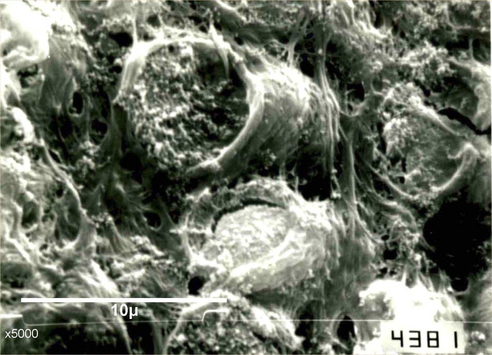
Mammals´ enamel is constituted in a 90% by hydroxyapatite crystals forming "prisms" and the interprismatic substance that surrounds them. Prisms present different shape according to the species. In some regions they are organized in parallel (Hunter-Schreger bands), and in others in a crisscross way. There is little information regarding the dog, so the aim of this study was to determine the ultramorphology of dog enamel by scanning electron microscopy (SEM). A total of 24 permanent healthy dog teeth were selected. They were cut with a diamond disc and the cut surface was etched with phosphoric acid. The teeth were processed to be studied with the SEM, and the prisms thickness was measured at different sites of the enamel. Dog prisms presented hexagonal shape, and an average thickness of 4,4 μm. Regions with Hunter-Schreger bands, and regions with cross prisms were found. In conclusion, prisms thickness of dog enamel does not vary according to the type of tooth or sector of the crown, except in the cusps where it is narrower. Prismatic organization was similar to that reported in previous dog enamel studies.
Downloads
References
Antonio Nanci A (2008) Ten Cate's Oral Histology: Development, Structure, and Function. Mosby Elsevier, St. Louis, MO, USA.
Chandra S (2004) Textbook of Dental and Oral Histology and Embryology with MCQs. Jaypee, New Delhi, India.
Fava M, Watanabe I, Moraes FF, Costa LRRS (1997) Prismless enamel in human non erupted deciduous molar teeth: A scanning electron microscopic study. Rev Odontol Univ Sa˜o Paulo 11:239–243.
Fava M, Myaki SI, Ramos CJ, Watanabe I (1999) Scanning electron microscopy observations of the prismless layer in fissures of erupted primary molars. Pós Grad Rev Fac Odontol São José dos Campos 2:1–7.
Garg N, Garg A (2015) Text book of operative dentistry, 3º ed. .Jaypee, New Delhi, India.
Gómez de Ferraris MA, Campos Muñoz A (2010) Histología, Embriología e Ingeniería Tisular Bucodental. Editorial Médica Panamericana, 3ºedición, Querétaro, México.
Gwinnett AJ (1992) Structure and composition of enamel. Oper Dent 5:10–17.
Hanaizumi Y, Kawano Y, Ohshima H, Hoshino M, Takeuchi K, Maeda T (1998) Three-dimensional Direction and Interrelationship of Prism in Cuspal and Cervical Enamel of Dog Tooth. Anat Rec 252: 355-368.
Hanaizumi Y, Yokota R, Domon T, Wakita M, Kozawa Y (2010) The initial process of enamel prism arrangement and its relationship to de Hunter-Schreger bands in dog teeth. Arch Histol Cytol 73 (1): 23-36.
Hueb De Menezes Oliveira MA, Torres CP, Gomez-Silva JM, Borsatto MC (2009) Microestructure and Mineral Composition of Dental Enamel of Permanent and Deciduous Teeth. Microsc Res Tech 73(5): 572-7.
Kuhar M, Cevc P, Schara M, Funduk N (1997) Enhanced permeability of acid etched or ground dental enamel. J Prosthet Dent 77:578–582.
Lynch ChD, Sullivan VRO, Dockery P, McGillycuddy CT, Sloan AJ (2010) Hunter-Schreger Band patterns in human tooth enamel. J Anat 217(2): 106-115.
Meckel AH, Griebstein WJ, Neal RJ (1965) Structure of mature human dental enamel as observed by electron microscopy. Arch. Oral Biol 10:775-78.
Osborn JW (1990) A 3-dimensional model to describe the relation between prism directions, parazones and diazones, and the Hunter- Schreger bands in human tooth enamel. Arch Oral Biol 35:869–878.
Osborn JW (1965) The nature of the Hunter-Schreger bands in enamel. Arch Oral Biol 10:929–935.
Rensberger JM (1993) Enamel microstructural specializations for strength in the teeth of Crocuta crocuta. Amer Zool 33: 102A (abstr).Skobe Z, Prostak KS, Trombly PL (1985) Scanning Electron Microscope Study of Cat and Dog Enamel Structure. J Morphol 184:195-203
Rowen D, Frandson W, Wilke L, Fails AD (2009) Anatomy and Physiology of Farm Animals, 7º ed. Wiley Blackwell, Singapur.
Sherwoo IA (2010) Essentials of operative dentistry. JAYPEE, New Delhi India.
Stefen C (1999) Enamel Microstructure of Recent and Fossil Canidae (Carnivora: Mammalia). J Vertebr Paleontol 19: 576-587.
Ten Cate AR (1989) Oral histology: development, structure and function, 3º ed. Mosby, St. Louis, MO, USA.
Tseng Z J (2011) Variation and Implications of Intra-Dentition Hunter-Schreger Band Pattern in Fossil Hyaenids and Canids (Carnivora, Mammalia) J Vertebr Paleontol 31(5):1163-1167.
Downloads
Published
How to Cite
Issue
Section
License
1. Política propuesta para revistas de acceso abierto
Los autores/as que publiquen en esta revista aceptan las siguientes condiciones:
- Los autores/as conservan los derechos de autor y ceden a la revista el derecho de la primera publicación, con el trabajo registrado con la licencia de atribución de Creative Commons, que permite a terceros utilizar lo publicado siempre que mencionen la autoría del trabajo y a la primera publicación en esta revista.
- Los autores/as pueden realizar otros acuerdos contractuales independientes y adicionales para la distribución no exclusiva de la versión del artículo publicado en esta revista (p. ej., incluirlo en un repositorio institucional o publicarlo en un libro) siempre que indiquen claramente que el trabajo se publicó por primera vez en esta revista.
- Se permite y recomienda a los autores/as a publicar su trabajo en Internet (por ejemplo en páginas institucionales o personales) despues del proceso de revisión y publicación, ya que puede conducir a intercambios productivos y a una mayor y más rápida difusión del trabajo publicado.

