¿Cómo generar un modelo tridimensional in vitro a partir de un adenocarcinoma mamario murino?
Mots-clés :
mamosferas, adenocarcinoma mamario murino, células madre mesenquimalesRésumé
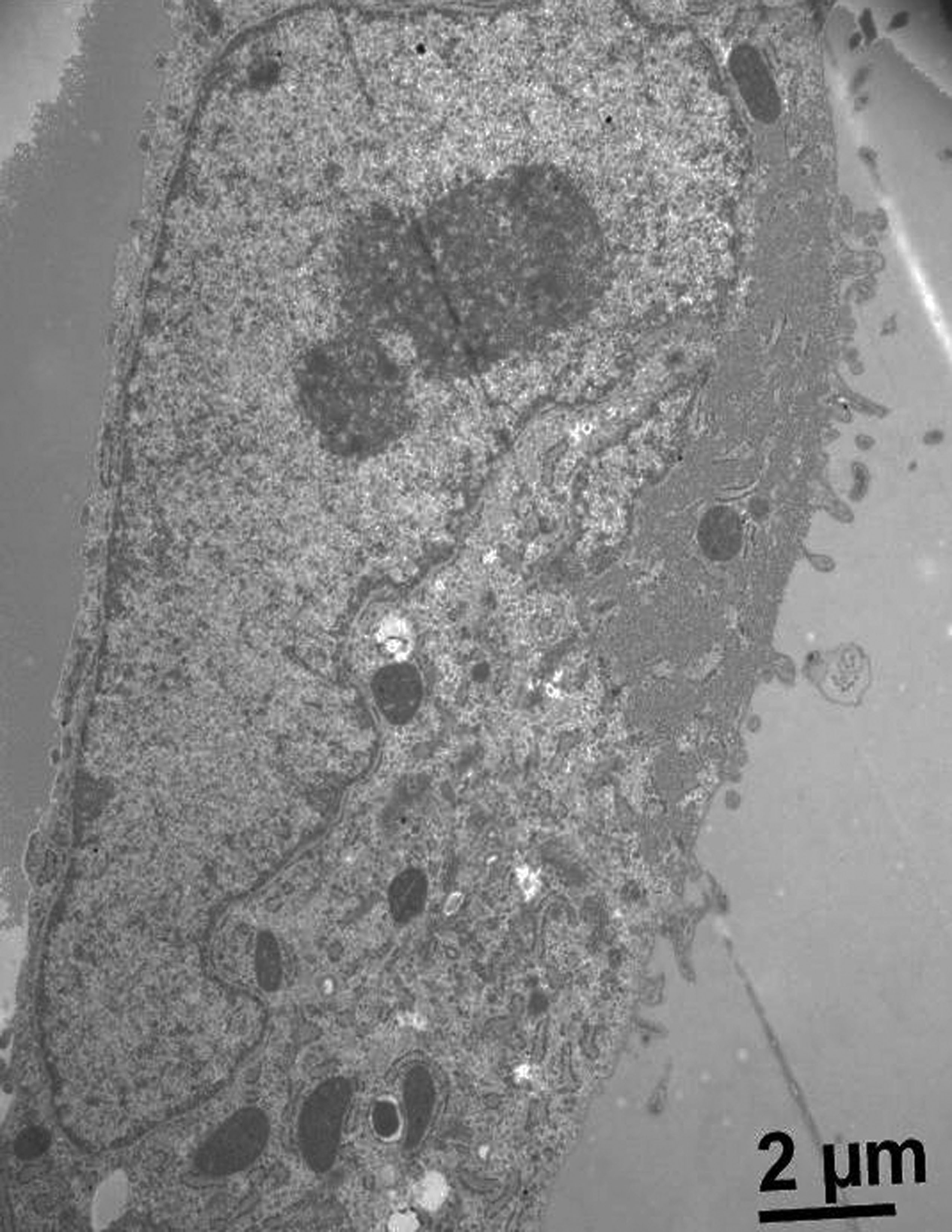
Las mamosferas son un modelo esferoide tumoral multicelular. Estas son utilizadas como un sistema de cultivo que se asemeja a la tridimensionalidad de los tumores. En este trabajo, hemos desarrollado este modelo utilizando un adenocarcinoma mamario murino con el fin de poder estudiar la fisiología tumoral y posibles aplicaciones terapéuticas. Se han probado diversas técnicas descriptas en trabajos previos. La mejor condición fue usar placas multiwells-96 en forma de V con la adición de una monocapa de agarosa, ya que se generaron esferas compactas y robustas. Además, hemos realizado tinciones histológicas de las mamosferas con hematoxilina-eosina para la observación de su estructura mediante microscopia óptica, y técnicas de inmunohistoquímica para evaluar el índice de proliferación celular y de neovascularización, con bromodeoxiuridina y VEGF (factor de crecimiento de endotelio vascular), respectivamente. Además, hemos estudiado la formación de estas mamosferas utilizando células tumorales y células madre mesenquimales derivadas de cordones umbilicales humanos para evaluar su interacción.
Références
Clarke KE, Tams DM, Henderson AP, Roger MF, Whiting A, Przyborski SA- (2016). A robust and reproducible human pluripotent stem cell derived model of neurite outgrowth in a three-dimensional culture system and its application to study neurite inhibition. Neurochem Int, 106:74-84.
Costa EC, Gaspar VM, Coutinho P, Correia IJ- (2014). Optimization of Liquid Overlay Technique to Formulate Heterogenic 3D Co-Cultures Models. Biotechnol. Bioeng. 11:1672-1685.
Cuiffo BG and Karnoub A- (2012). Mesenchymal stem cells in tumor development. Emerging roles and concepts. Cell Adh Migr, 6:220-230.
Fleisig H, Wong J- (2012). Measuring Cell Cycle Progression Kinetics with Metabolic Labeling and Flow Cytometry. J. Vis. Exp22:1-6.
Furnus CC, Inda AM, Andrini LB, García MN, García AL, Badrán A, Errecalde AL- (2003). Chronobiology of the proliferative events related to angiogenesis in mice liver regeneration after partial hepatectomy. Cell Biol Int 27: 383–386.García MN, Andrini LB, Inda AM, Ronderos JR, Hijano JC, Errecalde A- (2010). Changes in VEGF expression and DNA synthesis in hepatocytes from hepatectomized and tumour-bearing mice. Cell Biol. Int. 34:283–286.
García MN, Andrini L, Martinez M, Inda A, Palma MB, Miriuka S, Errecalde AL- (2015). Circadian rhythms of proliferation events in two mouse carcinomas. Biol- Rhythm Res. 46:573-578.
Hasselbach LA, Irtenkauf SM, Lemke NW, Nelson KK, Berezovsky AD, Carlton ET, Transou AD, Mikkelsen T, deCarvalho A- (2014). Optimization of High Grade Glioma Cell Culture from Surgical Specimens for Use in Clinically Relevant Animal Models and 3D Immunochemistry. J. Vis. Exp. 83:1-9.
Inda AM, García MN, Andrini LB, García AL, Fernández Blanco A, Furnus CC, Galletti SM, Brandoni J, Martinez JG, Prat GD, Errecalde AL- (2009). Evaluation of Angiogenesis with the Expression of VEGF-C and CD34 in Human Colon Cancer. Current Chemical Biology 3: 302-305.
Lee G, Hall R, Ahmed A- (2016). Cancer stem cells: celular plasticity, niche and its clinical relevance. J Stem Cell Res Ther. 6:1-20
Nagelkerke A, Bussink J, Sweep F, Span PN- (2013). Generation of multicellular tumor spheroids of breast cancer cells: How to go threedimensional. Anal Biochem 437: 17-19.
Razian G, Yu Y, Ungrin M- (2013). Production of large numbers of size-controlled tumor spheroids using microwell plates. J Vis Exp 81:1-6.
Solomon MA, Lemera J, D´Souza GG- (2016). Development of an in vitro tumor spheroid culture model amenable to high-throughput testing of potential anticancer nanotherapeutics. J Liposome Res. 26: 246-260.
Sun Z, Wang S, Chunhua Zhao R- (2014). The roles of mesenchymal stem cells in tumor inflamatory microenvironment. J Hematol Oncol. 7:2-10.
Suzuki K, Sun R, Origuchi M, Kanehira M, Takahata T, Itoh J, Umezawa A, Kijima H, Fukuda S, Saijo Y- (2011). Mesenchymal stromal cells promote tumor growth. Through the enhancement of neovascularization. Molmed. 17: 579-587.
Wharton´s jelly-derived multipotent mesenchymal stromal cells obtained from bovine umbilical cord and maintained in a defined serum-free three-dimensional system. BMC Biotechnology 12:1-11.
Wong C, Vosburgh E, Levine AJ, Cong L, Xu EY- (2012). Human neuroendocrine tumor cell lines as a three-dimensional model for the study of human neuroendocrine tumor therapy. J Vis Exp 66:1-7.
Téléchargements
Publié
Numéro
Rubrique
Licence
1. Política propuesta para revistas de acceso abierto
Los autores/as que publiquen en esta revista aceptan las siguientes condiciones:
- Los autores/as conservan los derechos de autor y ceden a la revista el derecho de la primera publicación, con el trabajo registrado con la licencia de atribución de Creative Commons, que permite a terceros utilizar lo publicado siempre que mencionen la autoría del trabajo y a la primera publicación en esta revista.
- Los autores/as pueden realizar otros acuerdos contractuales independientes y adicionales para la distribución no exclusiva de la versión del artículo publicado en esta revista (p. ej., incluirlo en un repositorio institucional o publicarlo en un libro) siempre que indiquen claramente que el trabajo se publicó por primera vez en esta revista.
- Se permite y recomienda a los autores/as a publicar su trabajo en Internet (por ejemplo en páginas institucionales o personales) despues del proceso de revisión y publicación, ya que puede conducir a intercambios productivos y a una mayor y más rápida difusión del trabajo publicado.

