Importancia del análisis microestructural del esmalte de la dentición decidua para la reconstrucción de la cronología del desarrollo dentario: una perspectiva antropológica
Mots-clés :
dentición decidua, esmalte, histología, marcadores de crecimiento, información documentalRésumé
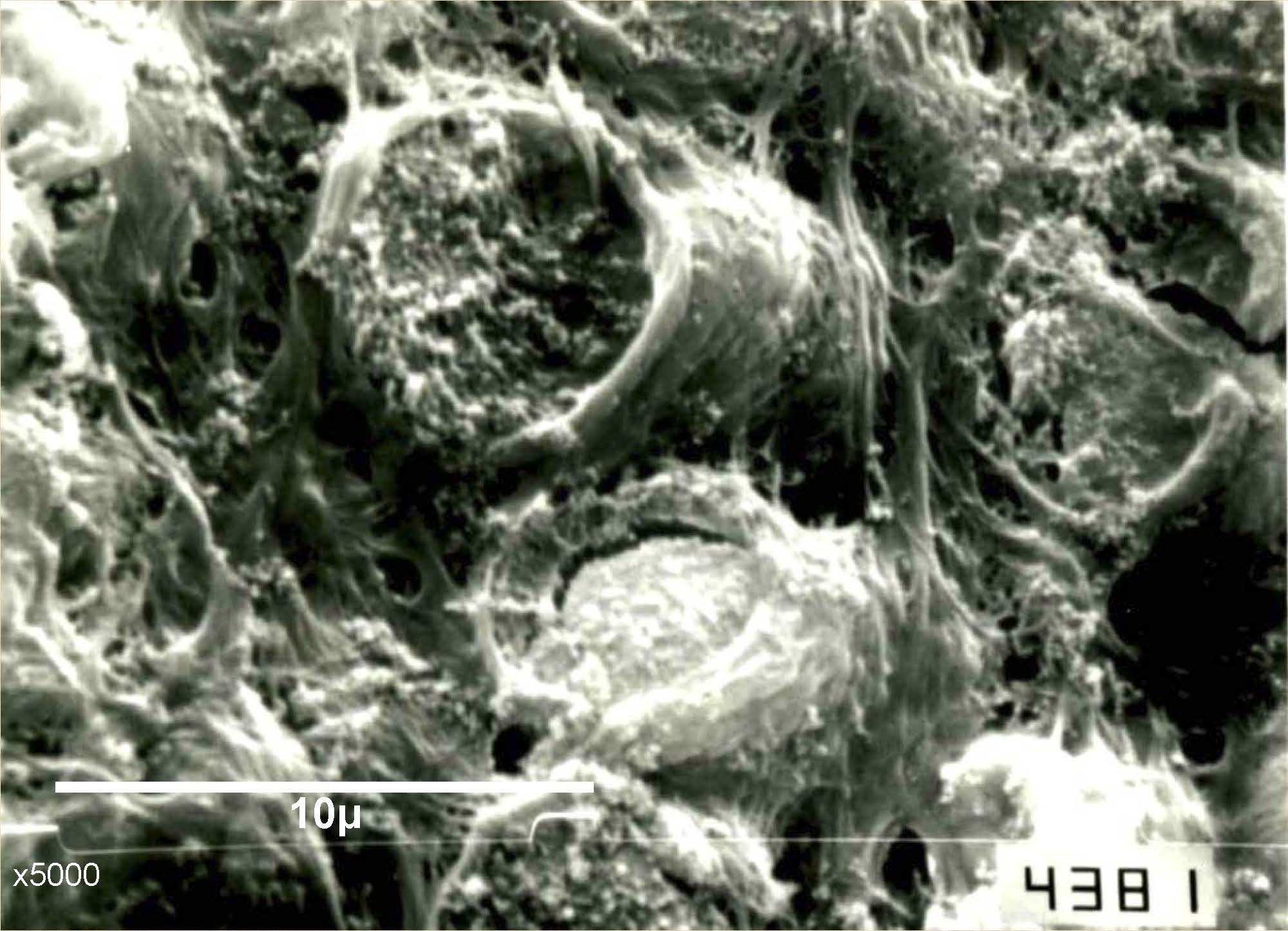
El proceso de mineralización de los dientes deciduos está definido por un ciclo en la actividad de secreción de las células formadoras del esmalte (ameloblastos), que alterna periodos de crecimiento, rápido y lento, y tiene como consecuencia la formación de bandas incrementales en el esmalte. Estos marcadores representan un registro temporal que puede ser una herramienta importante para el estudio del patrón y el tiempo de formación de las coronas dentarias, y de eventos de estrés ocurridos durante el crecimiento. El enfoque histológico ha sido aplicado sobre distintas muestras, tanto fósiles como arqueológicas y contemporáneas, brindando información sobre el crecimiento que no es posible obtener mediante otras técnicas. En los últimos años se han producido avances importantes en el campo de la histología que permitieron una aproximación más precisa a los procesos que regulan la formación de los tejidos duros del cuerpo, integrando información proveniente de distintas escalas de análisis. De esta manera, el presente trabajo tiene como objetivo revisar las principales premisas que subyacen al análisis de la microestructura dentaria en estudios antropológicos, la potencialidad y las problemáticas técnico-metodológicas de este enfoque, y finalmente, exponer líneas actuales de investigación en histología dentaria de individuos en crecimiento.
Références
Aka PS, Canturk N, Dagalp R, Yagan M (2009) Age determination from central incisors of fetuses and infants. Forensic Sci Int 184: 15-20.
AlQahtani SJ, Hector MP, Liversidge HM (2010) Brief communication: The London atlas of human tooth development and eruption. Am J Phys Anthropol 142: 481-490.
Antoine D, Hillson S, Dean MC (2009) The developmental clock of dental enamel: a test for the periodicity of prism cross-striations in modern humans and an evaluation of the most likely sources of error in histological studies of this kind. J Anat 214: 45-55.
Aranda C, Del Papa M (2009) Avances en las prácticas de conservación y manejo de restos humanos en Argentina. Rev Arg Antrop Biol 11: 89-94.
Bello SM, Thomann A, Signoli M, Dutour O, Andrews P (2006) Age and sex bias in the reconstruction of past population structures. Am J Phys Anthropol 129: 24-38.
Bernal V, Luna LH (2011) The development of dental research in Argentinean biological anthropology: current state and future perspectives. HOMO 62: 315-327.
Beynon AD, Clayton CB, Ramirez Rozzi FV, Reid DJ (1998) Radiographic and histological methodologies in estimating the chronology of crown development in modern humans and great apes: a review, with some applications for studies on juvenile hominids. J Hum Evol 35: 351-370.
Birch W, Dean MC (2014) A method of calculating human deciduous crown formation times and of estimating the chronological ages of stressful events occurring during deciduous enamel formation. J Forensic Leg Med 22: 127-144.
Bogin B, Smith BH (1996) Evolution of the human life cycle. Am J of Hum Biol 8: 703-716.
Bogin B (1999) Evolutionary perspective on human growth. Annu Rev Anthropol 28: 109-153.
Bollini G, Atencio JP, Luna L (2016) Caracterización de la dentición humana y aportes de la antropología dental para los estudios evolutivos, filogenéticos y adaptativos. En: Introducción a la Antropología Biológica, Madrigal L, González-José R (eds) http://scholarcommons.usf.edu/islac_alab_antropologia/1.
Boyde A (1963) Estimation of age at death of young human skeletal remains from incremental lines in dental enamel. En: Primate life history and evolution. Rousseau JD (ed) Wiley-Liss.
Bromage TG, Dean MC (1985) Re-evaluation of the age at death of immature fossil hominids. Nature 317: 525-527.
Bromage TG (1987) The biological and chronological maturation of early hominids. J Hum Evol 16: 257-272.
Bromage TG (1991) Enamel incremental periodicity in the pig-tailed macaque: a polychrome fluorescent labeling study of dental hard tissues. Am J Phys Anthropol 86: 205-214.
Bromage TG, Hogg RT, Lacruz RS, Hou C (2012) Primate enamel evinces long period biological timing and regulation of life history. J Theor Biol 305: 131-144.
Bromage TG, Lacruz RS, Hogg R, Goldman HM, McFarlin SC, Warshaw J, Dirks W, Perez-Ochoa A, Smolyar I, Enlow DH, Boyde A (2009) Lamellar bone is an incremental tissue reconciling enamel rhythms, body size, and organismal life history. Calcif Tissue Int 84: 388-404.
Canturk N, Atsu SS, Aka PS, Dagalp R (2014) Neonatal line on fetus and infant teeth: an indicator of live birth and mode of delivery. Early Hum Dev 90: 393-397.
Cardoso HF (2007) Accuracy of developing tooth length as an estimate of age in human skeletal remains: the deciduous dentition. Forensic Sci Int 172: 17-22.
Caropreso S, Bondioli L, Capannolo D, Cerroni L, Macchiarelli R, Condò SG (2000) Thin sections for hard tissue histology: a new procedure. J Microsc 199: 244-247.
Crowder C, Stout S (2011) Bone Histology: an anthropological perspective. Taylor & Francis.
Cui F-Z, Ge J (2007) New observations of the hierarchical structure of human enamel, from nanoscale to microscale. J Tissue Eng Regen Med 1: 185-191.
D´Addona LA, Gonzalez PN, Bernal V (2016) Variabilidad de las proporciones molares en poblaciones humanas: un abordaje empleando modelos del desarrollo y experimentales. Rev Arg Antropol Biol 18: 1-13.
de Boer HH, Aarents MJ, Maat GJR (2013) Manual for the preparation and staining of embedded natural dry bone tissue sections for microscopy. Int J Osteoarchaeol 23: 83-93.
Dean MC, Shellis RP (1998) Observations on stria morphology in the lateral enamel of Pongo, Hylobates and Proconsul teeth. J Hum Evol 35: 401-410.
Dean MC (2000) Incremental markings in enamel and dentine: what they can tell us about the way teeth grow. En: Function and evolution of teeth development, Teaford MF, Smith MM, Ferguson MWJ (eds) Cambridge University Press, Cambridge, New York, pp. 119-130.
Dean C (2000) Progress in understanding hominoid dental development. J Anat 197: 77-101.
Dean CM (2006) Tooth microstructure tracks the pace of human life-history evolution. Proc Biol Sci 273: 2799-2808.
Demirjian A, Goldstein H, Tanner JM (1973) A new system of dental age assessment. Hum Biol 45: 211-227.
Desántolo B, Inda AM (2016) Estimación microscópica de edad a partir de la zona cortical del fémur en individuos adultos: revisión metodológica. Rev Arg Antrop Biol 18: 1-12.
Dirks W (1998) Histological reconstruction of dental development and age at death in a juvenile gibbon (Hylobates lar). J Hum Evol 35: 411-425.
Eli I, Sarnat H, Talmi E (1989) Effect of the birth process on the neonatal line in primary tooth enamel. Pediatr Dent 11: 220-223.
Fava M, Watanabe IS, Fava de Moraes F, Costa LRdRSd (1997) Prismless enamel in human non-erupted deciduous molar teeth: a scanning electron microscopic study. Rev Odontol Univ São Paulo 11.
FitzGerald CM (1998) Do enamel microstructures have regular time dependency? Conclusions from the literature and a large-scale study. J Hum Evol 35: 371-386.
FitzGerald CM, Saunders SR (2005) Test of histological methods of determining chronology of accentuated striae in deciduous teeth. Am J Phys Anthropol 127: 277-290.
Fitzgerald CM, Rose JC (2008) Reading between the lines: dental development and subadult age assessment using the microstructural growth markers of teeth. En: Biological Anthropology of the human skeleton, Katzemberg MA y Saunders SR (eds) Wiley-Liss, New York, pp. 237-264.
FitzGerald C, Saunders S, Bondioli L, Macchiarelli R (2006) Health of infants in an Imperial Roman skeletal sample: perspective from dental microstructure. Am J Phys Anthropol 130: 179-189.
Fukuhara T (1959) Comparative anatomical studies of the growth lines in the enamel of mammalian teeth. Acta Anat Nipp 34: 322-332.
Garizoain G, Petrone S, García Mancuso R, Plischuk M, Desántolo B, Inda AM, Salceda SA (2016) Análisis de preservación ósea y dentaria en dos grupos etarios: su importancia en el estudio de conjuntos esqueléticos. Intersecciones Antropol 17: 353-362.
Gómez de Ferraris ME, Campos Muñoz A (2009) Histología, embriología e ingeniería tisular bucodental. Editorial Médica Panamericana.
Goodman AH, Rose JC (1990) Assessment of systemic physiological perturbations from dental enamel hypoplasias and associated histological structures. Am J Phys Anthropol 33: 59-110.
Gustafson G, Gustafson A (1967) Microanatomy and histochemistry of enamel. En: Structural and chemical organization of teeth, Miles AEW (ed) Academic Press, London, pp. 75-134.
Hastings MH (1991) Neuroendocrine rhythms. Pharmacol Ther 50: 35-71.
Hillson S (2005) Teeth. Cambridge University Press, Cambridge, New York.
Huda TFJ, Bowman JE (1995) Age determination from dental microstructure in juveniles. Am J Phys Anthropol 97: 135-150.
Irurita Olivares J, Alemán Aguilera I, Viciano Badal J, Luca S, Botella López M (2014) Evaluation of the maximum length of deciduous teeth for estimation of the age of infants and young children: proposal of new regression formulas. Int J Leg Med 128: 1354-1352.
Kavanagh KD, Evans AR, Jernvall J (2007) Predicting evolutionary patterns of mammalian teeth from development. Nature 449: 427-432.
Kodaka T, Sano T, Higashi S (1996) Structural and calcification patterns of the neonatal line in the enamel of human deciduous teeth. Scanning Microsc 10: 737-743.
Lacruz RS, Hacia JG, Bromage TG, Boyde A, Lei Y, Xu Y, Miller JD, Paine ML, Snead ML (2012) The circadian clock modulates enamel development. J Biol Rhythms 27: 237-245.
Le Cabec A, Tang N, Tafforeau P (2015) Accessing developmental information of fossil hominin teeth using new synchrotron microtomography-based visualization techniques of dental surfaces and interfaces. PloS one 10: e0123019.
Leigh SR (2001) Evolution of human growth. Evol Anthropol 10: 223-236.
Liversidge HM, Dean MC, Molleson T (1993) Increasing human tooth length between birth and 5.4 years. Am J Phys Anthropol 90: 307-313.
Luna L (2016) Some achievements and challenges of dental anthropology. ARC J Dent Sc 1: 5-9.
Lunt RC, Law DB (1974) A review of the chronology of calcification of deciduous teeth. J Am Dent Assoc 89: 599-606.
Manifold BM (2013) Differential preservation of children's bones and teeth recovered from early medieval cemeteries: possible influences for the forensic recovery of non-adult skeletal remains. Anthropol Rev 76: 23-49.
Mays S, Vincent S, Campbell G (2012) The value of sieving of grave soil in the recovery of human remains: an experimental study of poorly preserved archaeological inhumations. J Archaeol Sc 39: 3248-3254.
Mahoney P (2011) Human deciduous mandibular molar incremental enamel development. Am J Phys Anthropol 144: 204-214.
Mahoney P (2012) Incremental enamel development in modern human deciduous anterior teeth. Am J Phys Anthropol 147: 637-651.
Mahoney P (2013) Testing functional and morphological interpretations of enamel thickness along the deciduous tooth row in human children. Am J Phys Anthropol 151: 518-525.
Mahoney P (2015) Dental fast track: prenatal enamel growth, incisor eruption, and weaning in human infants. Am J Phys Anthropol 156: 407-421.
Mahoney P, Miszkiewicz JJ, Pitfield R, Schlecht SH, Deter C, Guatelli-Steinberg D (2016) Biorhythms, deciduous enamel thickness, and primary bone growth: a test of the Havers-Halberg Oscillation hypothesis. J Anat 228: 919-928.
Mann A, Lampl M, Monge J (1990) Patterns of ontogeny in human evolution: evidence from dental development. Am J Phys Anthropol 33: 111-150.
Marks MK, Rose JC, Davenport WD (1996) Technical note: thin section procedure for enamel histology. Am J Phys Anthropol 99: 493-498.
McFarlane G, Littleton J, Floyd B (2014) Estimating striae of Retzius periodicity nondestructively using partial counts of perikymata. Am J Phys Anthropol 154: 251-258.
Moorrees CFA, Fanning EA, Hunt EE (1963) Formation and resorption of three deciduous teeth in children. Am J Phys Anthropol 21: 205-213.
Moorrees CFA, Fanning EA, Hunt EE (1963) Age variation of formation stages for ten permanent teeth. J Dent Res 42: 1490-1502.
Nanci A (2008) Ten Cate's Oral Histology: Development, structure, and function. Mosby Elsevier.
Petrone S, Garizoain G, García M, Andrini L, García A, Inda AM (2016) Análisis microscópico del esmalte deciduo humano en distintos momentos del desarrollo. Rev Cs Morfol 18(1): 53-73.
Petrone S, Garizoain G (2017) Análisis histológico de esmalte dentario desde una perspectiva antropológica. Técnica de corte delgado para microscopía óptica. Cuadernos del INAPL SE 4(4): 108-116.
Ramirez Rozzi F (1996) Líneas de crecimiento en el esmalte dentario. Aplicación a los homínidos del Plio- Pleistoceno. Rev Arg Antrop Biol 1: 181-197.
Ramírez Rozzi F (1998) Can enamel microstructure be used to establish the presence of different species of Plio-Pleistocene hominids from Omo, Ethiopia?. J Hum Evol 35: 543-576.
Reid DJ, Dean MC (2006) Variation in modern human enamel formation times. J Hum Evol 50: 329-346.
Reid DJ, Beynon AD, Ramirez Rozzi FV (1998) Histological reconstruction of dental development in four individuals from a medieval site in Picardie, France. J Hum Evol 35: 463-477.
Risnes S (1998) Growth tracks in dental enamel. J Hum Evol 35: 331-350.
Sabel N, Johansson C, Kühnisch J, Robertson A, Steiniger F, Norén JG, Klingberg G, Nietzsche S (2008) Neonatal lines in the enamel of primary teeth: a morphological and scanning electron microscopic investigation. Arch Oral Biol 53: 954-963.
Saunders SR (2008) Juvenile skeletons and growth related studies. En: Biological anthropology of the human skeleton (2da edición), Katzemberg MA, Saunders S (eds) New Jersey: John Wiley & Sons, Inc pp. 117-147.
Seow WK (1986) Oral complications of premature birth. Aust Dent J 31: 23-29.
Seow WK, Young W, Tsang AK, Daley T (2005) A study of primary dental enamel from preterm and full- term children using light and scanning electron microscopy. Pediatr dent 27: 374-379.
Shellis RP (1998) Utilization of periodic markings in enamel to obtain information on tooth growth. J Hum Evol 35: 387-400.
Shinoda H (1984) Faithful records of biological rhythms in dental hard tissues. Chem Today 162: 34-40.
Simmer JP, Fincham AG (1995) Molecular mechanisms of dental enamel formation. Crit Rev Oral Biol Med 6: 84-108.
Smith BH (1986) Dental development in Australopithecus and early Homo. Nature 323: 327-330.
Smith BH (1992) Life history and the evolution of human maturation. Evol Anthropol 1: 134-142.
Smith TM (2006) Experimental determination of the periodicity of incremental features in enamel. J Anat 208: 99-113.
Smith TM (2008) Incremental dental development: methods and applications in hominoid evolutionary studies. J Hum Evol 54: 205-224.
Smith P, Avishai G (2005) The use of dental criteria for estimating postnatal survival in skeletal remains of infants. J Archaeol Sc 32: 83-89.
Smith TM, Tafforeau P, Le Cabec A, Bonnin A, Houssaye A, Pouech J, Moggi-Cecchi J, Manthi F, Ward C, Makaremi M (2015) Dental ontogeny in Pliocene and early Pleistocene hominins. PloS one 10: e0118118.
Tafforeau P, Boistel R, Boller E, Bravin A, Brunet M, Chaimanee Y, Cloetens P, Feist M, Hoszowska J, Jaeger J-J, Kay RF, Lazzari V, Marivaux L, Nel A, Nemoz C, Thibault X, Vignaud P, Zabler S (2006) Applications of X-ray synchrotron microtomography for non-destructive 3D studies of paleontological specimens. Applied Physics A 83: 195-202.
Tanevich A, Durso G, Batista S, Abal A, LLompart G, Llompart J, Martinez C, Licata L (2013) Microestructura del esmalte en dientes deciduos: los tipos de esmalte y la resistencia a la abrasión. UNR Journal 6: 1713-1718.
Teivens A, Mornstad H, Noren J, Gidlund E (1996) Enamel incremental lines as recorders for disease in infancy and their relation to the diagnosis of SIDS. Forensic Sci Int 81: 175-183.
Temple DH (2014) Plasticity and constraint in response to early-life stressors among late/final jomon period foragers from Japan: evidence for life history trade-offs from incremental microstructures of enamel. Am J Phys Anthropol 155: 537-545.
Tsutaya T, Yoneda M (2015) Reconstruction of breastfeeding and weaning practices using stable isotope and trace element analyses: a review. Am J Phys Anthropol 156: 2-21.
Żądzińska E, Lorkiewicz W, Kurek M, Borowska-Strugińska B (2015) Accentuated lines in the enamel of primary incisors from skeletal remains: a contribution to the explanation of early childhood mortality in a medieval population from Poland. Am J Phys Anthropol 157: 402-410.
Zanolli C, Bondioli L, Manni F, Rossi P, Macchiarelli R (2011) Gestation length, mode of delivery, and neonatal line-thickness variation. Hum Biol 83: 695-713.
Téléchargements
Publié
Numéro
Rubrique
Licence
1. Política propuesta para revistas de acceso abierto
Los autores/as que publiquen en esta revista aceptan las siguientes condiciones:
- Los autores/as conservan los derechos de autor y ceden a la revista el derecho de la primera publicación, con el trabajo registrado con la licencia de atribución de Creative Commons, que permite a terceros utilizar lo publicado siempre que mencionen la autoría del trabajo y a la primera publicación en esta revista.
- Los autores/as pueden realizar otros acuerdos contractuales independientes y adicionales para la distribución no exclusiva de la versión del artículo publicado en esta revista (p. ej., incluirlo en un repositorio institucional o publicarlo en un libro) siempre que indiquen claramente que el trabajo se publicó por primera vez en esta revista.
- Se permite y recomienda a los autores/as a publicar su trabajo en Internet (por ejemplo en páginas institucionales o personales) despues del proceso de revisión y publicación, ya que puede conducir a intercambios productivos y a una mayor y más rápida difusión del trabajo publicado.


