
Uso de la técnica de micromatrices de tejido en histología veterinaria
DOI:
https://doi.org/10.24215/15142590e008Palabras clave:
micromatrices de tejido, inmunohistoquímica, piel, caninosResumen
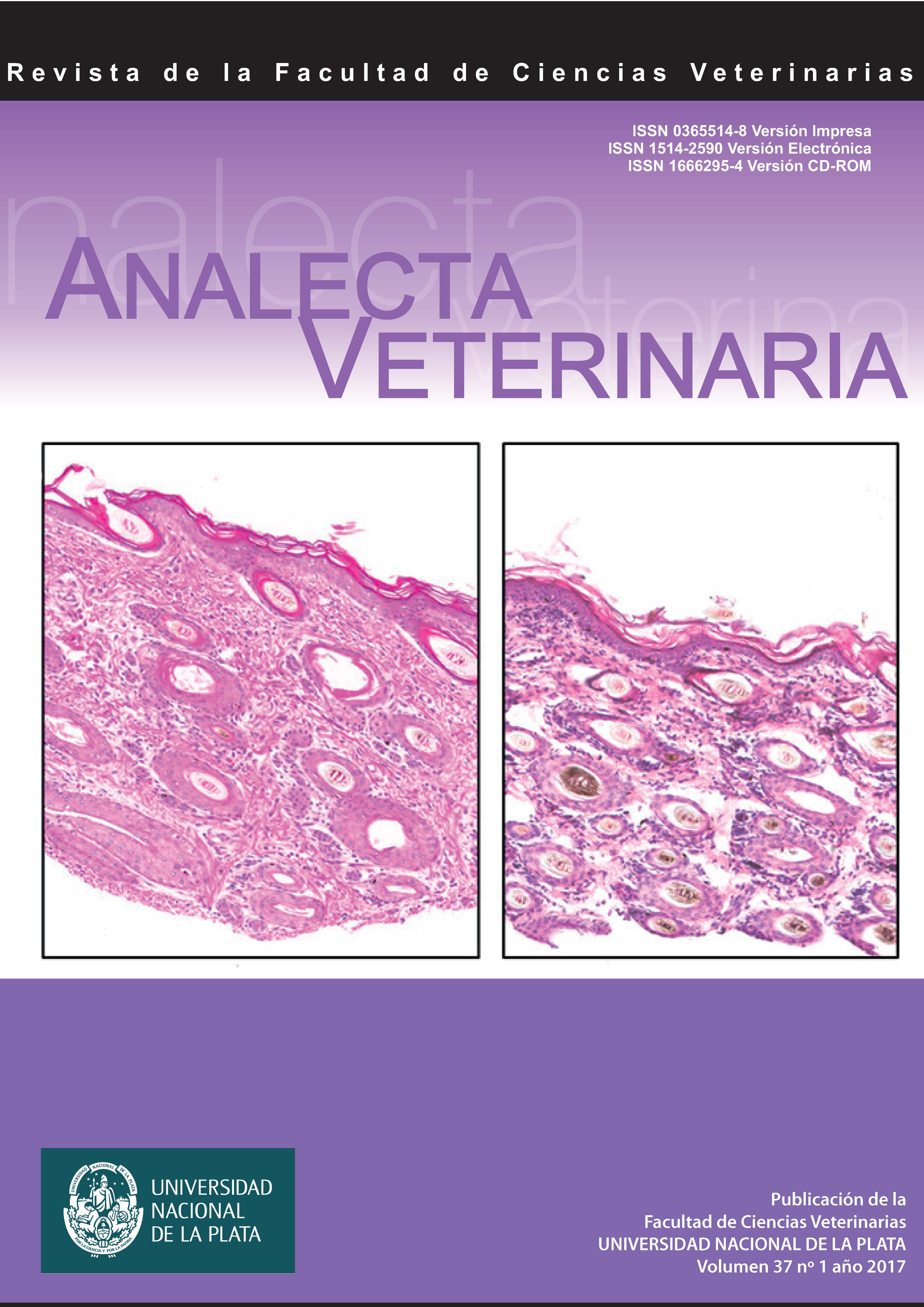
Las micromatrices de tejido son plataformas de alto rendimiento que permiten el análisis de decenas a cientos de muestras de tejidos en forma simultánea. Estas se han utilizado, en especial, para analizar tejidos neoplásicos. Sin embargo, los beneficios de su aplicación pueden ser aprovechados en otras áreas de investigación, como la embriología y la histología. El presente trabajo explora el uso de la técnica de micromatrices de tejido para el análisis morfológico y molecular de tejidos animales normales. Un total de 82 muestras de piel canina normal prenatal y posnatal fueron utilizadas para construir la matriz. A partir de ésta, se obtuvo una gran cantidad de secciones en las que se aplicaron técnicas de histología convencional y de inmunohistoquímica. Los resultados nos muestran la eficacia de la técnica para realizar el análisis morfológico y molecular de decenas de muestras de tejido normal en forma simultánea. Esto permite la evaluación más estandarizada de los tejidos, reduciendo así la variabilidad que puede ocurrir cuando se realizan ensayos sobre muestras individuales.
Descargas
Métricas
Descargas
Publicado
Cómo citar
Número
Sección
Licencia
Los autores/as conservan los derechos de autor y ceden a la revista el derecho de la primera publicación, con el trabajo registrado con la licencia de atribución de Creative Commons, que permite a terceros utilizar lo publicado siempre que mencionen la autoría del trabajo y a la primera publicación en esta revista.

Analecta Veterinaria por Facultad de Ciencias Veterinarias se distribuye bajo una Licencia Creative Commons Atribución-NoComercial-SinDerivar 4.0 Internacional.