La tomografía axial computarizada como herramienta para el diagnóstico y la planificación quirúrgica de la compresión medular
DOI:
https://doi.org/10.24215/15142590ep.%2039-44Keywords:
caninos, región toracolumbarAbstract
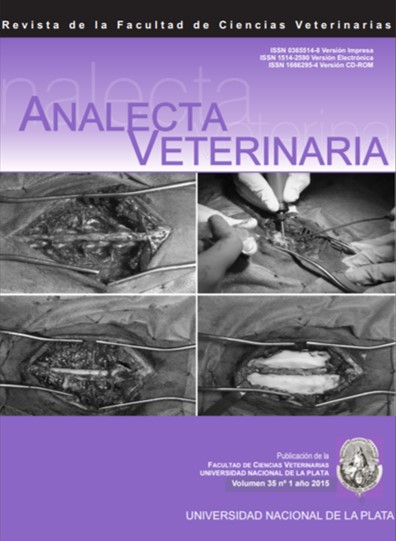
The herniated disc is the most common cause of spinal injury in dogs. In 85 % of cases it occurs at the thoracolumbar region. The Dachshund is one of the most susceptible breeds, due to inheritance. Anatomically, lesions are located mainly between the intervertebral spaces of T11-T12 to L1-L2. It is associated with a degeneration of the nucleus pulposus of the intervertebral disc, thus producing an extrusion or a protrusion. As a result of spinal cord compression the most frequent neurological disorders are included within the upper motor neuron syndrome (UMNS). A case of a 5 years old Dachshund female dog with kyphosis, thoracolumbar pain and a 15 days evolution paraparesis is described. To corroborate the diagnosis computerized axial tomography (CAT) was used. Images obtained assisted in defining the extent and nature of injuries, consisting of alterations in the appearance of the nucleus pulposus and a significant spinal cord compression. A decompressive surgery was proposed as a method to alleviate the neurological deficit. During surgery, images obtained by CAT were used to determine the number of vertebrae to be included in the laminectomy to facilitate removal of the extruded material and to more precisely locate the anatomic landmarks. Thirty days after surgery, the patient showed an incipient recovery of motor function and coordination of voluntary movements. It is concluded that neither loss of motor function nor severity of clinical signs allowed predicting the outcome of the case and the reversibility of the lesions. The use of CAT was an important tool for the diagnosis and resolution of the case.
Downloads
Metrics
Published
How to Cite
Issue
Section
License
Authors retain the copyright and assign to the journal the right of the first publication, with the with the terms of the Creative Commons attribution license. This type of license allows other people to download the work and share it, as long as credit is granted for the authorship, but does not allow them to be changed in any way or used them commercially.

Analecta Veterinaria by School of Veterinary Sciences, National University of La Plata is distributed under a Creative Commons Attribution-NonCommercial-NoDeriv 4.0 International License.


