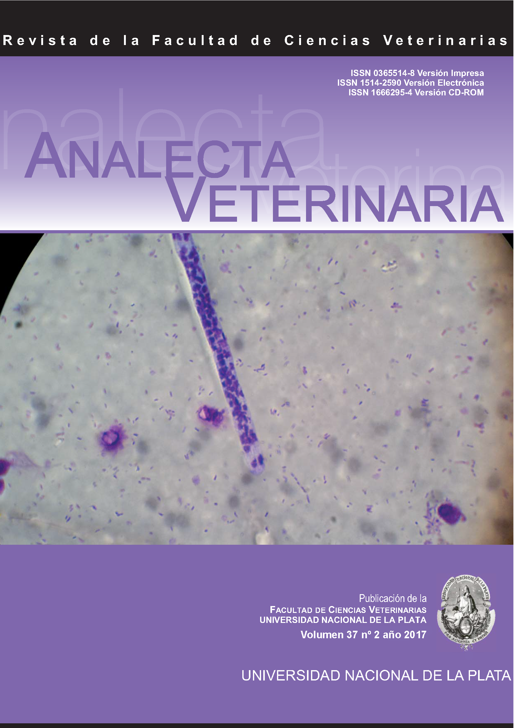
Morphology of Rangelia vitalii parasitic stages in samples from naturally infected dogs
DOI:
https://doi.org/10.24215/15142590e017Keywords:
Rangelia vitalii, morphology, diagnosisAbstract
The rangeliosis is a tick-borne disease affecting domestic and wild canids caused by Rangelia vitalii and transmitted by the tick Amblyomma aureolatum. The disease has been diagnosed in southern Brazil since the beginning of XX century, and recently in Argentina and Uruguay which could be related to an increase of the distribution area of the parasite and susceptible hosts. The parasite is an intracellular apicomplexan protozoon closely related with Babesia spp. in phylogenetical studies, however it showed differences in the life cycle. Parasitic stages have been observed in monocytes, erythrocytes and neutrophils, and in endothelial cells from several organs as well as free on plasma. The present study describes the morphology of 4 different parasitic stages observed in blood samples from naturally infected dogs in Argentina.
Downloads
Metrics
Downloads
Published
How to Cite
Issue
Section
License
Authors retain the copyright and assign to the journal the right of the first publication, with the with the terms of the Creative Commons attribution license. This type of license allows other people to download the work and share it, as long as credit is granted for the authorship, but does not allow them to be changed in any way or used them commercially.

Analecta Veterinaria by School of Veterinary Sciences, National University of La Plata is distributed under a Creative Commons Attribution-NonCommercial-NoDeriv 4.0 International License.


